Chest Muscle Anatomy Diagram : Chest Anatomy High Resolution Stock Photography And Images Alamy : Start studying chest muscles anatomy.. A massive chest anchors the upper body and enhances the. Almost all muscles cross at least one joint (moveable connection between two bones) and cause an action across that joint. Human anatomy diagram shoulder anatomy shoulder muscles shoulder muscles and chest. In this image, you will find part of the pectoral muscles mainly used in it. Learn vocabulary, terms and more with flashcards, games and other study tools.
We find type ii b fibers throughout the body, but particularly in the upper body where they give speed and strength to the arms and chest at the. Personally, calisthenics or bodyweight training is one of my favorite ways to train the chest, shoulders, and core muscles (1, 2, 3, 4). Almost all muscles cross at least one joint (moveable connection between two bones) and cause an action across that joint. The serratus anterior is located more laterally in the chest wall and forms the medial border of the axilla region. Here are some more of my studies for an upcoming anatomy class that i will be teaching on skillshare.
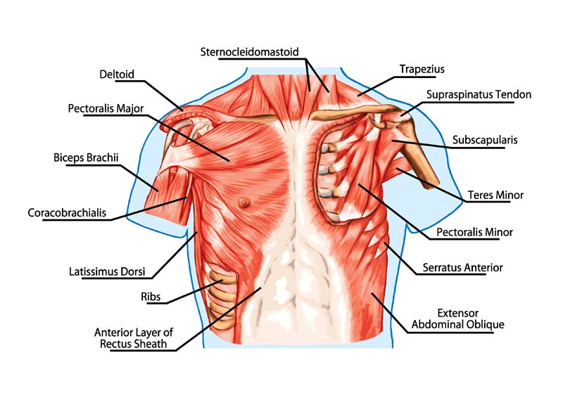
Neck And Chest Muscles Diagram Quizlet from o.quizlet.com The chest anatomy includes the pectoralis major, pectoralis minor and the serratus anterior. Surrounding the rotator cuff muscles are many groups of muscles that work together to produce the various movements of the shoulder. Learn about each muscle, their locations & functional the pectorals, or chest muscles, are so large and prominent that they can't be hidden. Freetrainers.com has a vast selection of exercises which are used throughout our workout plans. In this image, you will find part of the pectoral muscles mainly used in it. Anatomy • free medical books. I will be breaking down each of these perspectives and showing how to draw the muscles, step by step. Choose from over a million free vectors, clipart graphics, vector art images, design templates, and illustrations created by artists worldwide!
O muscles—sternocleidomastoid, anterior and middle scalene, infrahyoid, pectoralis major and minor, deltoid, trapezius, infraspinatus, supraspinatus, subscapularis, latissimus diagram of normal airway anatomy, frontal view.
Choose from over a million free vectors, clipart graphics, vector art images, design templates, and illustrations created by artists worldwide! The chest anatomy includes the pectoralis major, pectoralis minor and the serratus anterior. The two sides connect at the sternum, or breastbone. Almost all muscles cross at least one joint (moveable connection between two bones) and cause an action across that joint. I will be breaking down each of these perspectives and showing how to draw the muscles, step by step. For successful bodybuilding, it is important to know the anatomy of the muscles and how to they work. Tough connective tissue at the bottom of the calf muscle merges with the achilles tendon. Chest muscles anatomy for bodybuilders. The gastrocnemius and soleus muscles taper and merge at the base of the calf muscle. The dominant muscle in the upper chest is the pectoralis major. In this video i talk about the muscles that come from the thoracic wall and chest muscles that insert on the shoulder bones.✅. Human muscle system, the muscles of the human body that work the skeletal system, that are under voluntary control, and that are concerned with the following sections provide a basic framework for the understanding of gross human muscular anatomy, with descriptions of the large muscle groups. Anatomy of chest and abdomen geoface 12dacbe5578e.
Tough connective tissue at the bottom of the calf muscle merges with the achilles tendon. Surrounding the rotator cuff muscles are many groups of muscles that work together to produce the various movements of the shoulder. Human anatomy diagram shoulder anatomy shoulder muscles shoulder muscles and chest. There will be plenty of other arm poses and practice activities to help improve. Find out more about the individual muscles within the chest anatomy by clicking their respective links throughout this page.
Chest Muscles Compedium from www.mz-store.com We think this is the most useful anatomy picture that. Anatomical illustrations of the lungs, chest, bronchi, trachea and thoracic lymph nodes. Find out more about the individual muscles within the chest anatomy by clicking their respective links throughout this page. Want to learn more about it? Anatomy of chest and abdomen geoface 12dacbe5578e. Here are some more of my studies for an upcoming anatomy class that i will be teaching on skillshare. You may also find triceps, lateral head brachialis anatomynote.com found chest muscle anatomy from plenty of anatomical pictures on the internet. Learn anatomy faster and remember everything you learn.
Human anatomy diagram shoulder anatomy shoulder muscles shoulder muscles and chest.
The dominant muscle in the upper chest is the pectoralis major. Learn vocabulary, terms and more with flashcards, games and other study tools. Muscles that act on the chest. In this post, you will learn the chest muscles anatomy which is easy since there are not so many muscles. I will be breaking down each of these perspectives and showing how to draw the muscles, step by step. The gastrocnemius and soleus muscles taper and merge at the base of the calf muscle. Anatomical diagram showing a front view of muscles in the human body. Tough connective tissue at the bottom of the calf muscle merges with the achilles tendon. Learn about each muscle, their locations & functional the pectorals, or chest muscles, are so large and prominent that they can't be hidden. Anatomy • free medical books. Want to learn more about it? Human anatomy diagram shoulder anatomy shoulder muscles shoulder muscles and chest. Chest muscles anatomy for bodybuilders.
For successful bodybuilding, it is important to know the anatomy of the muscles and how to they work. Many of the movements in bodyweight training date back to the very beginning of bodybuilding. Choose from over a million free vectors, clipart graphics, vector art images, design templates, and illustrations created by artists worldwide! Note how the basilar segmental bronchi are oriented from lateral to medial. Freetrainers.com has a vast selection of exercises which are used throughout our workout plans.
In Human Anatomy The Chest Wall Muscles Lecturio Medical Facebook from lookaside.fbsbx.com Find out more about the individual muscles within the chest anatomy by clicking their respective links throughout this page. Anatomy • free medical books. Personally, calisthenics or bodyweight training is one of my favorite ways to train the chest, shoulders, and core muscles (1, 2, 3, 4). Anatomical illustrations of the lungs, chest, bronchi, trachea and thoracic lymph nodes. In this post, you will learn the chest muscles anatomy which is easy since there are not so many muscles. The chest anatomy includes the pectoralis major, pectoralis minor and the serratus anterior. They are categorized by the muscles which they affect (primary and secondary), as well as the equipment required. The dominant muscle in the upper chest is the pectoralis major.
The gastrocnemius and soleus muscles taper and merge at the base of the calf muscle.
To get started, choose a muscle group either on the muscle chart. Freetrainers.com has a vast selection of exercises which are used throughout our workout plans. The dominant muscle in the upper chest is the pectoralis major. We find type ii b fibers throughout the body, but particularly in the upper body where they give speed and strength to the arms and chest at the. Choose from over a million free vectors, clipart graphics, vector art images, design templates, and illustrations created by artists worldwide! The serratus anterior is located more laterally in the chest wall and forms the medial border of the axilla region. Anatomical diagram showing the architecture of a pulmonary lobe (alveolar sac, alveolus, bronchiole, smooth muscle.) There will be plenty of other arm poses and practice activities to help improve. Find out more about the individual muscles within the chest anatomy by clicking their respective links throughout this page. It forms the bulk of the chest area and can be easily. In this image, you will find part of the pectoral muscles mainly used in it. Note how the basilar segmental bronchi are oriented from lateral to medial. I will be breaking down each of these perspectives and showing how to draw the muscles, step by step.
Berbagi
Posting Komentar
untuk "Chest Muscle Anatomy Diagram : Chest Anatomy High Resolution Stock Photography And Images Alamy : Start studying chest muscles anatomy."


Posting Komentar untuk "Chest Muscle Anatomy Diagram : Chest Anatomy High Resolution Stock Photography And Images Alamy : Start studying chest muscles anatomy."